
Overbite before and after side view
A side view can show how orthodontic treatment changes upper incisor overlap, tooth angulation, and the relationship between the jaws.
The short answer
A side view can show how orthodontic treatment changes upper incisor overlap, tooth angulation, and the relationship between the jaws. A photograph alone cannot prove the bite has been corrected, because the clinician also checks the teeth from the front, the chewing contacts, and imaging when indicated.
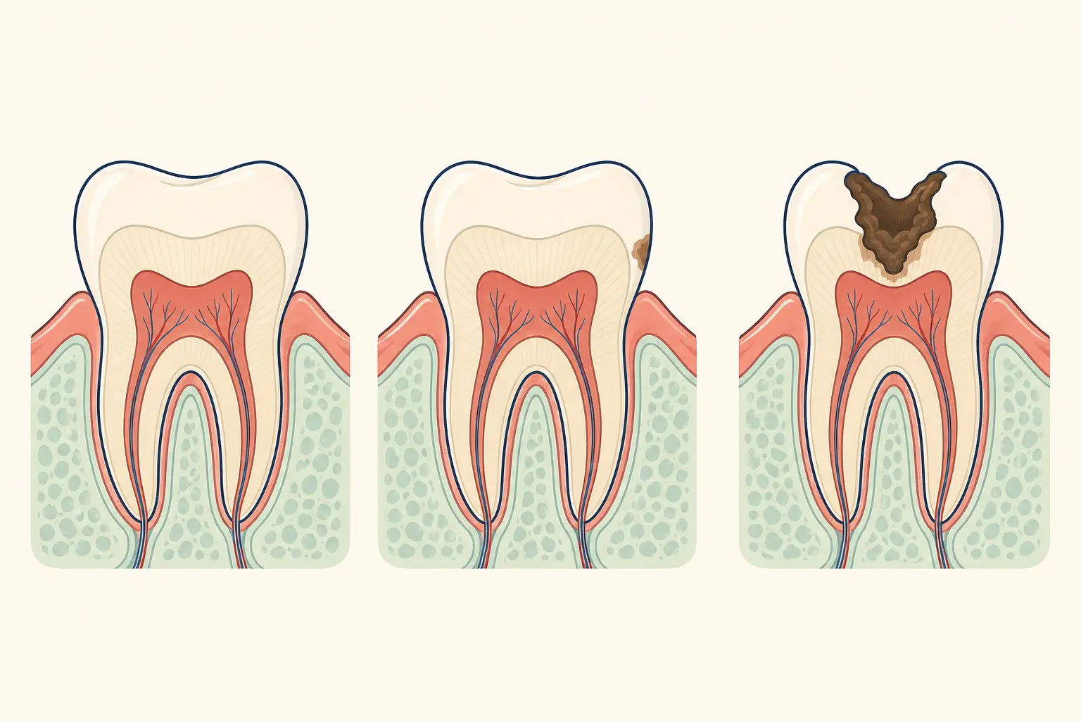
How is the overlap between your front teeth defined?
An overbite is the vertical overlap of the upper front teeth over the lower front teeth when the teeth are together. A profile photograph may show a deep bite, protruding incisors, or a lower jaw position, but it cannot separate tooth movement from growth, head posture, or camera angle.
How should you compare overbite photographs accurately?
Bring consistent before and after photographs to an orthodontic assessment and compare the same side, head position, lighting, and relaxed lip posture. The orthodontist records the overbite, overjet, tooth positions, periodontal condition, and jaw relationship, then discusses braces, clear aligners, growth guidance, or other treatment when appropriate.
When can an overbite cause dental problems?
Arrange an assessment when the lower incisors contact the palate, the gums are traumatised, the front teeth cannot meet comfortably, or chewing is difficult. Seek prompt care for facial injury, a suddenly altered bite, or a loose or displaced tooth.
Does your overbite mainly involve teeth or jaw structure?
Ask how much of the overbite comes from tooth position and how much comes from jaw structure. Ask which records will be used, what the treatment target is, how retention will work, and whether the side-view change reflects a stable bite rather than appearance alone.
Frequently asked questions
Can a side photograph diagnose an overbite?
It can suggest a facial or incisor pattern, but diagnosis requires an intraoral bite examination and sometimes additional records.
Why can the profile look different after braces?
Tooth angulation, incisor position, lip support, and bite relationships can change, while the visible result also depends on posture and photography.
Keep reading
Who wrote this page
This library is published by Dental Nation, a licensed dental group practising in the United Arab Emirates. Pages are written and overseen by the same clinical team that treats patients in our clinics, and are held to a fixed editorial standard for safety and accuracy.
This page is patient education, not medical advice, and cannot replace an examination. Anything urgent — facial swelling, trauma, uncontrolled bleeding or severe pain — needs a dentist or emergency care now.