
Causes of gingival pigmentation
Gingival pigmentation is usually caused by melanin, but medication, tobacco, inflammation, or a local lesion can also change gum colour.
The short answer
Gum colour varies naturally because melanocytes in the gingival epithelium produce melanin. Brown or grey areas may be even on both sides, or limited to one site when a local factor is involved.
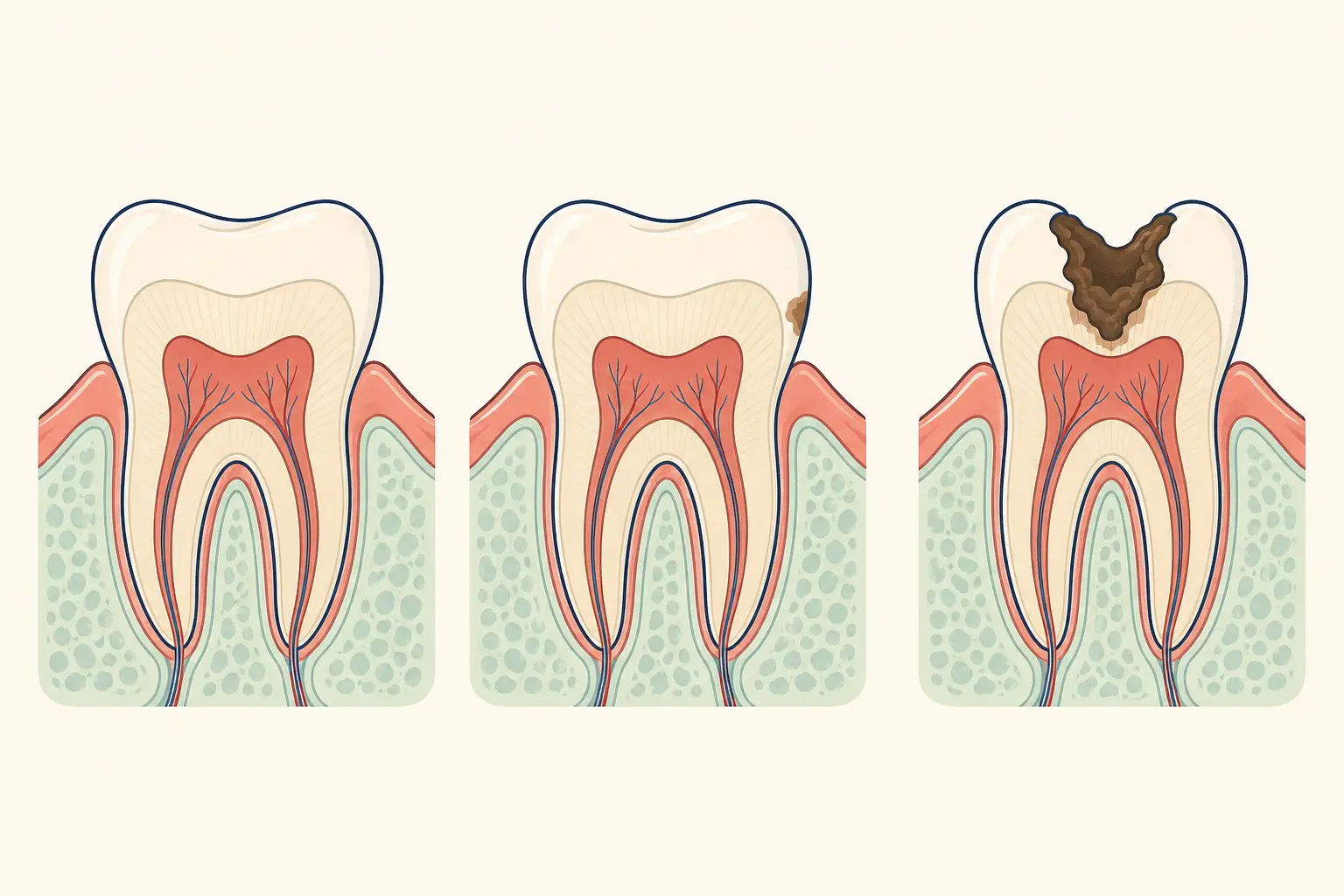
How does a dentist assess the pattern of gum pigmentation?
A dentist examines the pattern, surface, bleeding, medicines, tobacco exposure, and recent changes. Stable, symmetrical colour often needs observation, while a new or irregular patch may need photographs, periodontal testing, or a biopsy.
What changes in gum pigmentation should you have examined?
Arrange an examination for pigmentation that is new, enlarging, uneven, raised, ulcerated, bleeding, painful, or associated with a neck lump. Seek prompt care if a dark area changes quickly or does not heal.
When could gum pigmentation need prompt dental assessment?
Ask whether the colour pattern is normal melanin, whether a medicine or tobacco could contribute, and whether examination, monitoring, or tissue sampling is appropriate.
Frequently asked questions
Can this problem have more than one cause?
Yes. Dental findings often reflect several factors, so the examination must link the symptom or change to its cause.
What happens at the first visit?
The dentist reviews your history, examines the relevant teeth and gums, and selects tests that clarify the diagnosis.
Keep reading
Who wrote this page
This library is published by Dental Nation, a licensed dental group practising in the United Arab Emirates. Pages are written and overseen by the same clinical team that treats patients in our clinics, and are held to a fixed editorial standard for safety and accuracy.
This page is patient education, not medical advice, and cannot replace an examination. Anything urgent — facial swelling, trauma, uncontrolled bleeding or severe pain — needs a dentist or emergency care now.