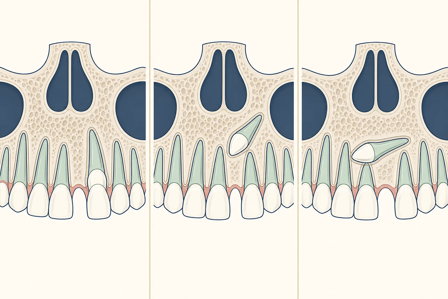
Impaction can occur when there is not enough space, the tooth follows an unusual path, or nearby teeth affect its eruption. Developmental and inherited factors can contribute to the pattern.
The useful clinical stage is based on whether the relevant teeth are visible, partly erupted, or still within the jaw, and whether nearby teeth or the bite are affected. Imaging can clarify position when the finding cannot be seen directly.
A late-erupting canine, a retained baby tooth, crowding, or movement of nearby teeth makes assessment more important. Imaging helps show the tooth’s position and its relationship to roots of adjacent teeth.
Diagnosis combines dental history, examination, and imaging when needed. The clinician assesses tooth development, spacing, gums, bite, and the relationship between the affected area and nearby teeth before discussing a plan.
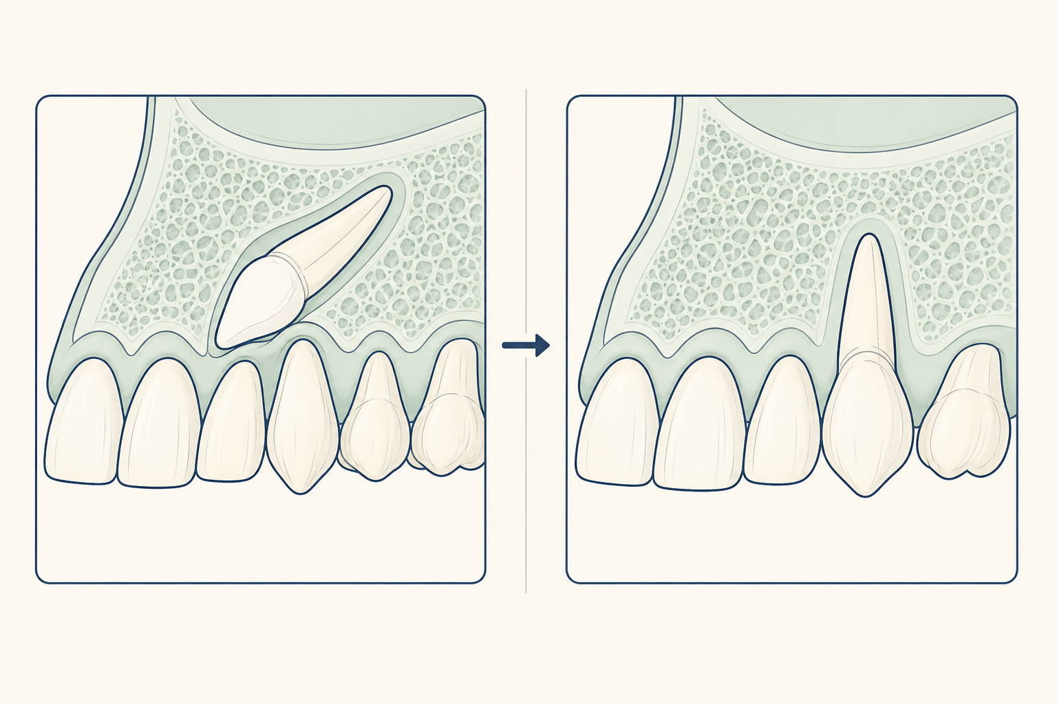
Options can include observation, creating space, exposing and guiding the tooth into position, or removal when keeping the tooth is not suitable. The plan depends on position, root development, and the surrounding teeth.
Without assessment, the issue may affect eruption, tooth position, tooth structure, comfort, or the bite, depending on the condition. A planned review makes it possible to act if those findings change.
Some eruption problems cannot be prevented. Routine checks during the mixed-dentition years can identify a canine that is late or off course and allow referral when needed.
An impacted canine is located with examination and imaging, then managed according to its position, the developing bite, and its effect on nearby teeth.