
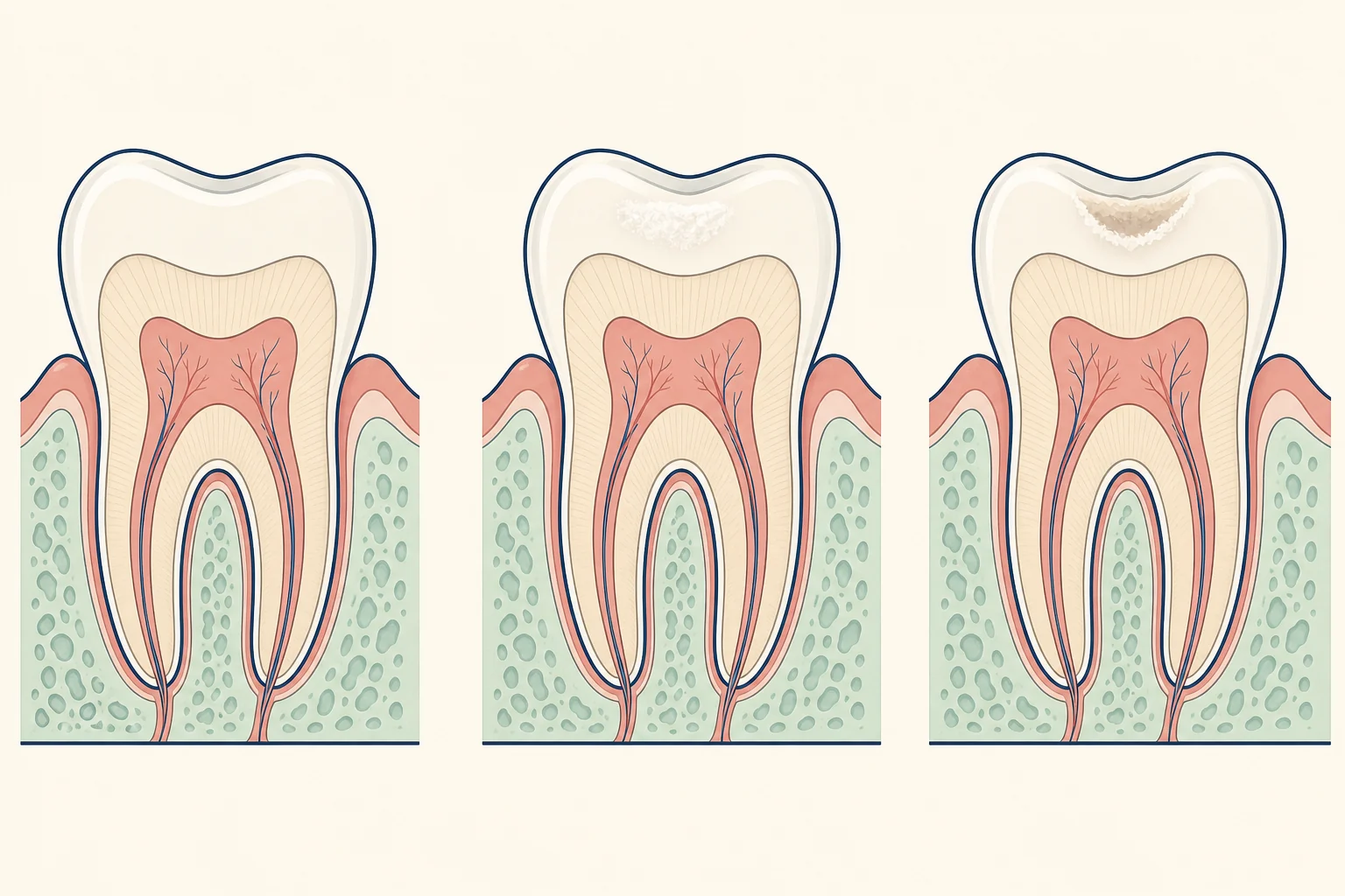
What the change looks like
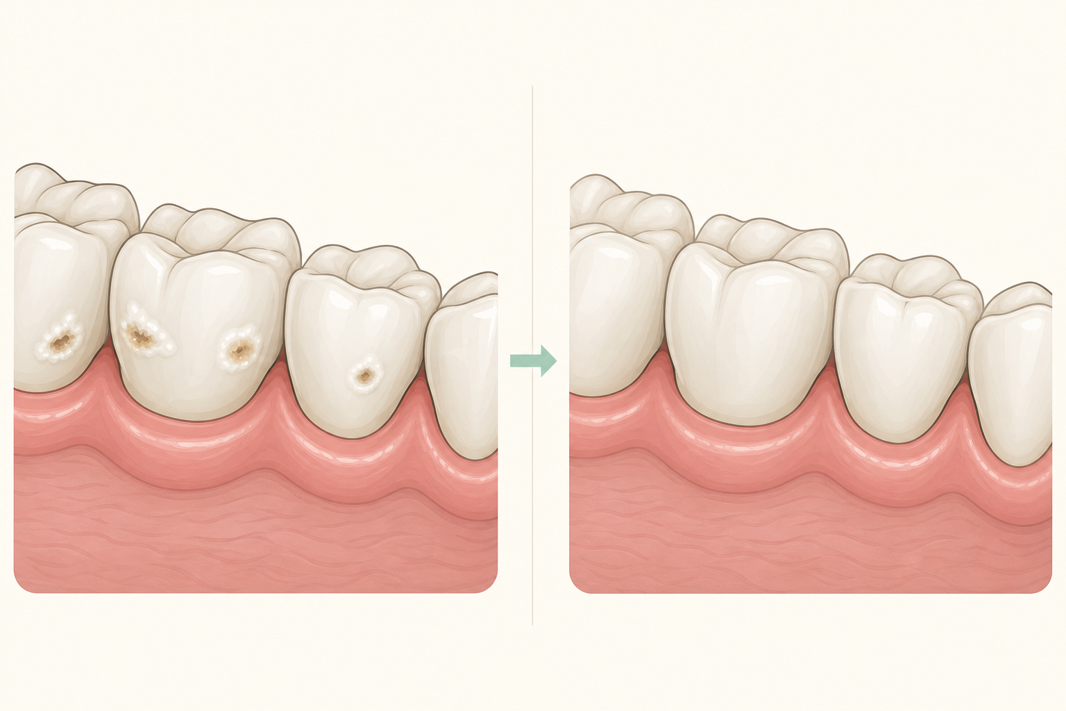
What might enamel caries feel or look like?
With continued acid exposure, smooth enamel can become rough, then develop microcavities and cavitation. Lesions vary, but some remain in enamel on radiographs for three to four years.
[2]How does decay begin in enamel?
Frequent sugar intake can shift plaque bacteria toward acid production. Acid removes mineral below the enamel surface; when enough mineral is lost, the white spot becomes visible.
[2]What does an enamel-stage finding mean?
Early enamel change corresponds to ICDAS 1 or 2 and can be arrested or reversed with preventive measures. Greater surface porosity can create enamel microcavities, corresponding to ICDAS 3.
[2]What can raise the chance of further decay?
Regular exposure to sugar and a cariogenic biofilm drive enamel mineral loss. Keeping plaque undisturbed allows that acid challenge to continue.
[2]How is enamel caries checked?
Assessment records the surface appearance and whether the lesion is active. Bitewing radiographs can show how far a lesion extends, including lesions that look non-cavitated clinically.
[2]What care may be discussed for an enamel lesion?
An accessible lesion that can be kept clean may not need a filling. Remineralisation, biofilm removal and sealing are non-invasive options; cavitated lesions can be controlled with minimally invasive care.
[2][3]| Clinical finding | Care discussion |
|---|---|
| An enamel lesion assessed as suitable for prevention | Preventive measures and follow-up may be considered.[2][3] |
| A lesion needing restorative management | The dentist can explain the reason for restoration and the tooth structure involved.[2][3] |
What should I ask about costs in Dubai?
Why should enamel caries be assessed?
How can I lower the chance of new caries?
Frequently asked questions
Can enamel caries be found before a cavity is obvious?
Yes. A clinical examination can identify a caries lesion and assess its activity and extent before deciding on care.
[2]