
Central Giant Cell Granuloma
Central giant cell granuloma is a benign jaw neoplasm that may be silent at first, then expand and affect bone, teeth, or the jaw cortex.
[1][2]The short answer
Central giant cell granuloma is often silent at the beginning. It later becomes expansile, and a larger lesion may produce a sharply defined radiolucency, displace tooth roots, or leave a tooth without enough bony support. Larger jaw lesions can be multilocular, and more aggressive lesions may breach the cortex.
[1][2]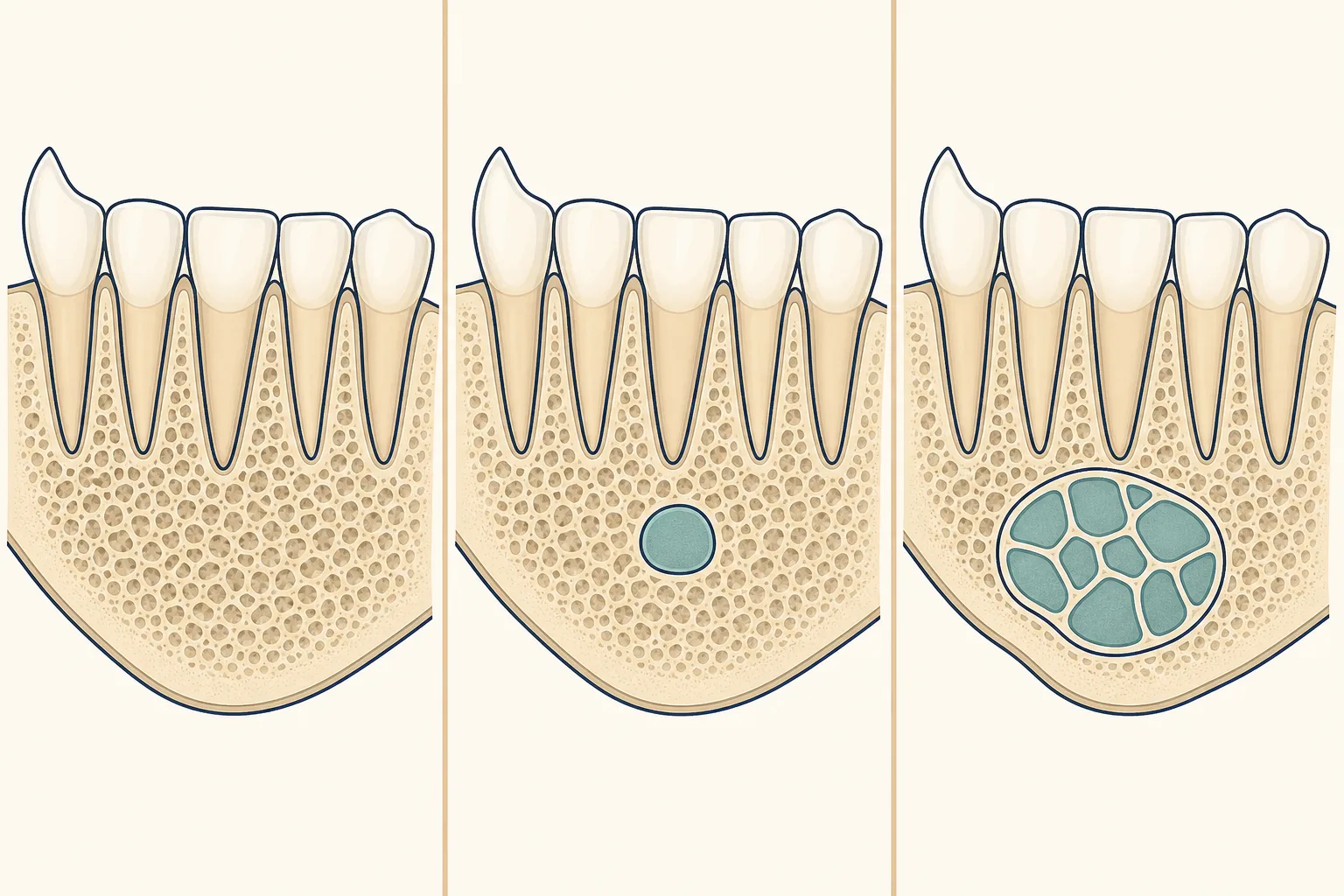
What the change looks like
What symptoms can central giant cell granuloma cause?
What causes central giant cell granuloma?
The lesion is linked with a proliferation of fibroblasts and multinucleated giant cells, but its pathogenicity remains uncertain. Multiple cases have been reported with mutations in the RAS/MAPK pathway, especially in syndromal cases. The lesion's possible relationship to long-bone tumours and some syndromes is one reason accurate classification matters.
[1][2]How can the lesion progress?
Who is more often affected?
How is central giant cell granuloma diagnosed?
Diagnosis requires clinicopathological correlation with radiological findings and, sometimes, serum evaluation. The lesion must be distinguished from other jaw lesions with overlapping histology, including central giant cell tumour, because confusing the two can lead to unnecessarily mutilating surgery. Radiographs may show a sharply demarcated radiolucency, a sclerotic margin, or faint calcifications.
[1][2]What treatment is used for different lesion patterns?
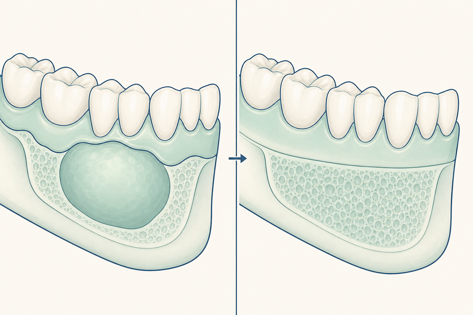
Curettage is the first-line treatment and is almost always curative. For large or aggressive lesions, particularly in a growing facial skeleton where curettage could be mutilating, calcitonin and injections of steroids into the lesion have been attempted. Interferon-alpha-2A has also been suggested as an additional treatment.
[1][2]| Lesion pattern | Treatment described |
|---|---|
| Typical lesion | Curettage is the first-line choice and is almost always curative.[1][2] |
| Large or aggressive lesion | Medical treatment with calcitonin or intralesional steroids has been attempted when curettage may be mutilating.[1][2] |
What shapes treatment planning in Dubai?
The main planning distinction is whether curettage is suitable or whether the lesion is large or aggressive enough that another approach has been attempted. A Dubai treatment discussion therefore needs to account for the lesion's size, behaviour, and the possibility of close clinical and radiographic follow-up after treatment.
[1][2]What problems can the lesion cause?
An expanding lesion can displace tooth roots, reduce the bony support around a tooth, or become large enough to form multiple compartments. In more aggressive cases, the cortex may be breached. The diagnostic distinction from a central giant cell tumour also matters because the wrong classification can lead to unnecessarily destructive surgery.
[1][2]